Features of the anatomy of nails on the fingers and toes. Nail care and maintenance. Nail diseases.
Nails are derivatives of the epidermis, dense plates on the tips of human fingers and toes (however, all primates have them). Despite the fact that they are devoid of nerve endings and do not hurt, marigolds act as indicators of the condition of the entire body.
Firstly, in order to look beautiful and have a stylish manicure, and secondly, in order to understand in time what is wrong with your health, you need to be aware of the anatomy and functions of the nail plates on the hands and feet.
Structure and functions of nails
Nails are a unique part of the human body. Their anatomy is quite complex, but by studying it you can learn some interesting facts.
Animal claws and human nails have very similar anatomy.
- The closest “relatives” of the nails on a person’s fingers and toes are his own hair, as well as animal hooves
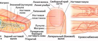
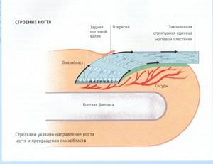
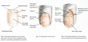
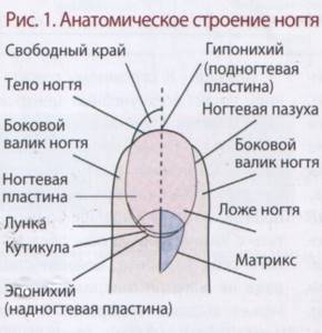
- The horny protective plate consists of three parts: the root (other names are matrix, matrix), the body and the free edge. The root is formed by living epidermal cells, and the body and free edge are dead
- The root part of the nail, hidden under the skin, in the nail fissure. We can not see. But its dimensions are not small, they make up a third of the visible part. The white semicircle visible on the plate near the lower roller is a continuation of the matrix. It is called lunula
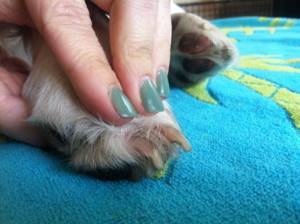
- The body of the nail rests on the nail bed. The average length of this part of the keratinized plate on the hands is 1.5 cm, width – 1 cm, thickness – 0.7 mm. On the feet, the extreme phalanges and, accordingly, the nail plates on the first and the remaining four fingers differ significantly in size, while the thickness of the plate on the big toe is approximately 1 mm
- The horny plate itself, naturally, is devoid of blood vessels. But there are extremely many of them under it, in the nail bed. It is these vessels that provide nutrition to the nail.
- Between the plate and the bed there is a thin layer of living cells, hyponychium
- Rollers are folds of skin located at the bottom and sides of the nail body. They are connected to the horny plate by the cuticle.
- The matrix consists of living epithelial cells - onychoblasts. They feed very intensively on blood, constantly divide and become horny, forming the protein keratin, which makes up the dead part of the plate
- The matrix is responsible for how the visible part of the nail looks - its shape, thickness, strength, growth rate, smoothness, etc. Injuries to the nail root directly affect the appearance of the plate
- The growth rate of the nail on the finger is up to 4 mm per month, on the toe – up to 3 mm during the same time. Interestingly, the growth process occurs faster in women. Also, nails are cut more often in the summer.
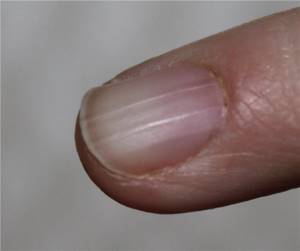
- The body of the nail, although represented by dead cells, is dense, shiny, elastic, and has a pleasant pink tint, if, of course, the person is healthy. This is explained by the fact that between keratin contains sulfur atoms (cysteine), between its parts in the plate there are “gaskets” of fat and water. The pink color of the plate is given by the blood circulating in the blood vessels located underneath it.
- The free edge of the nail can grow as much as it is strong and elastic, and as much as the person wants. A classic manicure is characterized by a length of 2 to 5 mm. Stiletto nails with an unusual design can be longer. As it grows, the free edge of the nail plate curls and takes the form of a spiral.
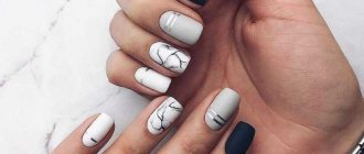
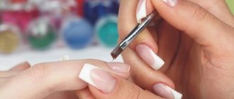
- Manicurists give the free edge of the nail various shapes by filing.
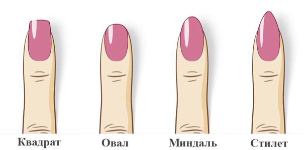
Classic shapes of the free edge of the nail.
IMPORTANT: There is a separate official science that studies the anatomy and functions of nails, and also diagnoses their condition. It is called "onychology"
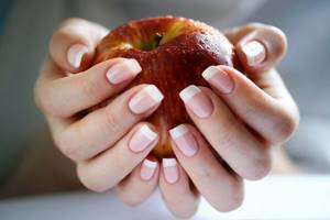
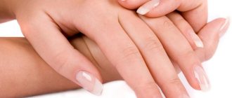
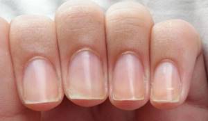
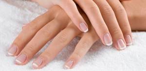
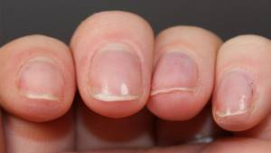
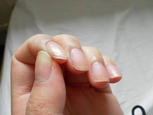
Healthy, shiny, flexible, strong nails are needed to protect your fingertips.
The main function of nails is to protect the outer phalanges of the fingers from the negative effects of environmental factors, in particular mechanical, chemical, vibration, temperature, etc. Also, marigolds:
- Needed to itch
- Help a person manipulate various objects, giving the fingertips the necessary hardness
- Help a person tactilely evaluate an object
- They are a means of self-expression
Yes, thanks to the possibilities of modern nail design, nails for a woman are a decoration comparable to clothing, accessories, and jewelry. The ability to take care of nails is also a valuable quality for a man.
The singer Countess has a record long nails - 91 cm.
How to speed up nail growth using home methods
By speeding up the process of cell division in the matrix, you can speed up the growth of the plates. You shouldn't expect magic, but good results can be obtained from the following procedures:
- Daily massage of the fingertips and matrix with soft circular movements or with the index and middle finger of the other hand closed in a lock.
Hand massage: types, technique, recommendations for the procedure - Taking vitamins and minerals for growth will speed up the process a little and make the plates less brittle.
As a result, there will be a faster growth effect, so the ends will not flake and break off. Vitamins for nails - which ones are needed and how they affect - Application of special varnishes and serums to accelerate growth.
Particular attention should be paid to the matrix zone, since this is where cell division occurs. How to use IBX System, all features of the product line - Protect the tips during any household work and physical activity so that they do not simply grind off on hard objects.
- Oil and water based baths with the addition of any components that improve blood circulation in the growth area.
For example, citrus oils, rosemary and tea tree. Hand baths: 30 recipes for different purposes - Capsules with an oily solution of vitamins A and E. You can directly rub the composition into the matrix area 2 times a week for 5-7 weeks under plastic gloves for 30 minutes.
What does the structure of a fingernail look like: diagram
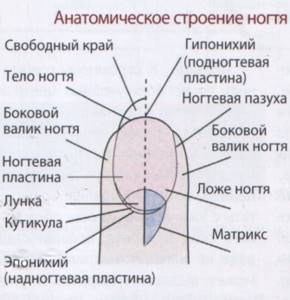
Diagram of the structure of a fingernail.
Detailed diagram of the structure of the nail.
Nail and nail plate.
general information
The nail is a regularly renewed tissue. And the structure of the nail is interesting mainly to women and, rather, only from the point of view of manicure and selection of varnish. In fact, knowing the structure of the nail plate is of great importance not only for beauty, but also for the health of nails. For greater clarity, below are the main reasons why this knowledge is so relevant in our time for any person who cares about their appearance.
- Firstly, understanding how the nail is structured and how it grows will help improve your daily care and avoid nail problems;
- Secondly, knowing the structure of nails is a prerequisite for many modern professions, for example, nail service specialists. After all, only a fully developed professional can perform a safe manicure and pedicure;
- Thirdly, even the most primitive diagnosis and treatment of nails is impossible without this knowledge.
Thus, having learned the structure of the nail, a person will improve his knowledge of his own body and will understand how best to keep it in shape. This will allow you to create the perfect manicure, possible only on healthy plates.
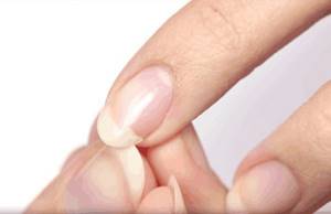
Cuticle on the nail - anatomy
From the ridges around the body of the nail, a thin protective film, called the cuticle, seems to grow onto the horny plate itself.
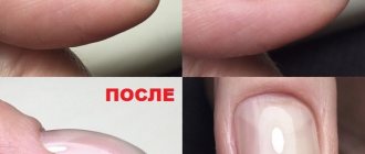
IMPORTANT: The cuticle consists of two types of cells - living and dead. Living cells are located closer to the skin folds, dead cells are located closer to the keratin plate. The dead part of the cuticle is easily damaged and develops hangnails, which quite often leads to inflammation of the living areas of the skin around the nail
The cuticle performs a protective function.
The main function of the cuticle is protective. The film is needed to prevent bacteria, dust, and other foreign bodies from getting into the cracks between the nail and the skin. Onychologists and manicurists are still debating whether it is necessary to remove the cuticle.
Previously, it was believed that a beautiful manicure with this film was simply impossible, and it was mercilessly cut off with scissors or bitten off with tweezers. Today, the majority is inclined to believe that the living part of the cuticle is needed. After softening with special means, the dead material is removed using a router attachment or carefully pushed away with a stick. It is also recommended to care for the cuticle using:
- baths
- massage
- special oils and creams
Theory of connection between fingers and internal organs: diagram
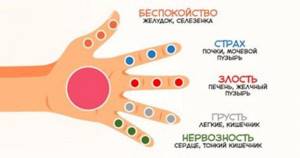
Our body is a single system and everything in it is interconnected. Some believe that fingers and toes are connected to our internal organs and reflect their condition. Massage therapists and some other specialists use this knowledge in their work.
If you notice any changes on your nails, knowing which finger belongs to which organ, you will be able to determine in which organ a malfunction may have occurred.
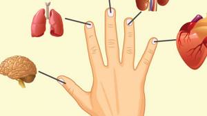
- Little finger – Heart
- Unnamed – Kidneys
- Middle – Intestines
- Index – Lungs
- Big – Brain
Remember, no video lessons will teach a manicurist the correct tactics of behavior when a client has problem nails. Moreover, if you come for a medical manicure, pay attention to the specialist’s diplomas - these are some of the most important documents that give him the legal and moral right to carry out his activities!
Be healthy and beautiful!
Author Evgenia Lizarenko especially for the website Style40plus.ru
Nail structure and manicure
To work in the nail industry, you must, in addition to having the desire and ability, complete courses and receive a certificate.
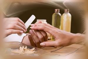
A manicurist must know the anatomy of the nail.
There are a huge number of manicure and pedicure courses in any city. Regardless of their cost and duration, the first thing future nail designers will have to become familiar with is the anatomy of the nail, its injuries and diseases. They need this block of knowledge to:
- understand what exactly they will have to work with
- protect clients from injuries and damage during manipulation of nails, and protect yourself from the troubles that follow these injuries and damage
- answer numerous questions that clients have
IMPORTANT: By the way, every person who does their own manicure should have knowledge of the structural features of nails. After all, it is ignorance that causes lasting injuries to the plate, matrix, skin, and infection during inept care procedures. Often similar troubles occur in children who get manicures from their parents.
Here are a few questions that manicurists hear especially often:
- Why are my nails so short? This parameter is determined genetically. The matrix is responsible for the shape of the plate. But the length of the nail plates is also influenced by external factors. For example, parents “disfigure” children’s marigolds when they damage the hyponychium in infancy by cutting them too short. Short nails are also common among those who have a habit of biting them.
- My nails feel thick. But why do they often break? The fact is that nail plates are very hygroscopic. Having absorbed water, they thicken, but lose elasticity, and, accordingly, break. It is recommended to wear special gloves for frequent contact with water.
- My nails are growing too slowly. Why? The speed of nail growth, again, is determined by heredity. Also, it depends on hormones. For example, pregnant women quickly acquire long nails. To increase your growth rate, you need to: eat properly and sufficiently; be in the sun; take vitamins; massage your fingers (this promotes more active blood supply to the matrix); be examined for the presence of cardiovascular and endocrine diseases, treat them if necessary
- The nails are dead. Why does it hurt me to trim or file them? It’s not the nails that hurt, because they don’t have nerves. Pain occurs when the well-innervated hyponychium is injured. This occurs when the nail beds are cut too short. Or, on the contrary, the nails have been long for too long, and the hyponychium has grown over them. To eliminate discomfort, it is recommended to lubricate the inner surface of the nail plate with oil and gently remove the skin that has grown on it using an orange stick.
- I hurt my nail and it turned black. What will happen now? You can read in detail about nail bruises in the article: link
What the changes will tell you: description and photo
Surprisingly, our nails are able to tell us about the changes that occur deep in our body.
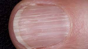
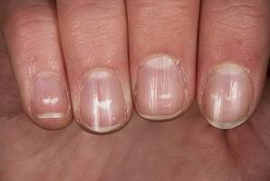
- • Longitudinal stripes – lack of vitamins, disruption of the gastrointestinal tract;
- • Transverse stripes - the cause of occurrence is a strict diet, or mechanical damage to the matrix (including manicure instruments);
- • Concave nail plate – indicate problems with the endocrine system;
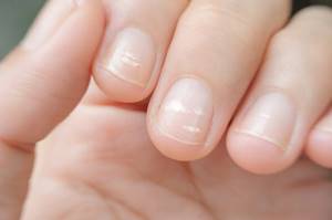
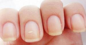
- • White spots - they appear due to the formation of air between the layers of the nail plate. Depending on the intensity, they indicate a lack of zinc, nervous tension, heart failure;
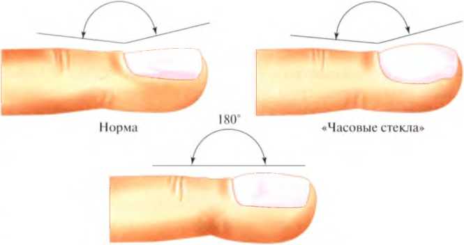
- • The effect of watch glasses is a sign of serious diseases of the heart, lungs, blood vessels, and gastrointestinal tract.
- • Severe, unfounded fragility is a sign of a serious vitamin deficiency that needs to be replenished.
- • Change in color of the nail plate
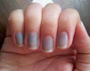
- – blue – indicates poor blood supply to both the nails and, possibly, the whole body. You shouldn’t joke about this, as oxygen deficiency can also manifest itself, which is very dangerous to health;
- – yellowness – speaks of intoxication of the body, that is, a failure of the organs that cleanse our body: kidneys, gall bladder, liver, sometimes lungs;
- – with a green tint – indicate purulent inflammation;
- – white – this is the first sign of liver cirrhosis. If the whitishness is not very pronounced, it may be a sign of a disease of the gastrointestinal tract and heart. If only half of the nail plate is white, pay attention to the condition of the kidneys.
- – purple color – indicates that the body is overloaded and is working with all its might. The main reason for this condition is blood stagnation associated with heavy foods that predominate in the diet.
- – general pallor – indicates anemia or problems with the spleen.
- • Indentations in the form of small dots are a sign of psoriasis or eczema. May sometimes indicate arthritis;
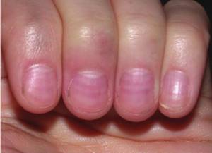
- • Lack of lunula – lack of B vitamins and anemia;
- • Large lunulae – problems with the heart, blood vessels and blood pressure;
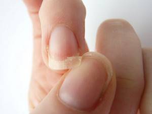
- • Exfoliating – notify about an unbalanced diet and lack of vitamins;
- • The nail plate moves away from the nail bed. This usually occurs due to mechanical damage, however, the cause can also be: allergies, fungus, various dermatological diseases, diseases of the nervous system and others. In this case, it is better to consult a doctor, because the symptom is associated with a huge number of disorders.
- • Black stripes – if the nail has not been injured (and you are unlikely to forget this), you need to pay attention to the health of the gastrointestinal tract.
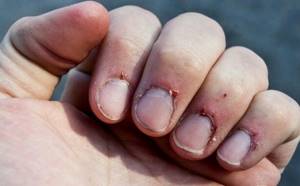
- • Hangnails – lack of vitamins.
An uneven nail plate and other deviations from the norm can be symptoms of a variety of diseases, so be sure to consult a doctor.
Pedicure basics and nail structure
The toes are subject to extreme stress due to walking upright and wearing shoes. When doing a pedicure, you need to think not only about aesthetics, but also about the health of your feet.
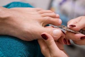
You can only do a pedicure correctly if you know the structure of the nail.
Here are some rules:
- It is better to push back the cuticles on the toes rather than cut them off
- The nails on the four small fingers can be cut short, at the root. On a large one, the length of the free edge should be about 1 mm
- When shortening the nail on your big toe, which is thick, you need to be very careful so as not to break it. If the nail has grown too much, it is trimmed; if it grows moderately, it is filed down. Shorten the free edge from the corners, not from the middle
- The corners of the toenails should not be rounded, otherwise they may grow into the skin.
IMPORTANT: If any injury to the nail or skin around it occurs during the pedicure procedure, you must use an antiseptic. There is a high risk of developing an infectious process
Nail sinuses (sinuses).
Nail sinuses are depressions at the transition points from the nail fold to the nail plate. The nail sinuses are protected from the penetration of bacteria and dirt by the cuticle, although this protection is not always enough, therefore, when performing a manicure, the sinuses are carefully processed with a manicure instrument to remove ptegirium and other deposits. Treatment of the nail sinuses is very important for maintaining the healthy condition of the entire nail apparatus and, especially, for preparing for subsequent coating with varnishes, gels and acrylics, preventing peeling of the artificial coating.
Nail structure and diseases
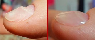
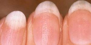
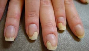
A healthy nail is strong, elastic, smooth, translucent, and has a pleasant color. Fragility, yellowness, dullness, furrows and irregularities indicate a particular disease. Brittle nails are caused by:
- lack of nutrition (deficiency of proteins and vitamins)
- the destructive effects of water, household chemicals, and other external factors
- prolonged exposure to the sun
- typing, playing musical instruments
- habit of peeling off varnish
- onychomycosis (fungal nail disease)
Brittle nails.
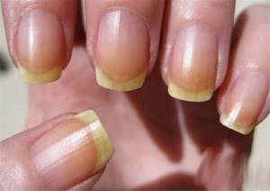
The nail plate becomes dull and yellow due to:
- smoking
- cardiovascular, endocrine diseases
- taking certain medications
- elderly
- fungus
Irregularities and grooves on the nail plate appear if:
- the root of the nail is injured
- unbalanced human nutrition
- a person has iron deficiency anemia
Furrows on the nails.
Layering of nails is explained:
- using household chemicals without gloves
- lack of vitamins
- habit of biting your nails or picking off polish
- allergies
- internal diseases
- nail fungus
Peeling nails.
About nail fungus and methods of treating it is written here: link
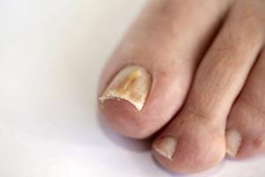
Toenail fungus.
Causes of injury to the free edge of the nail
It is easily damaged and delaminates even with minimal mechanical impact. The causes of trauma to the free edge of the nail are often fungal infections, solar ultraviolet radiation, and violation of cosmetic treatment technology.
The nail apparatus has a special property called immune privilege. It weakens some inflammatory processes and enhances protective reactions.
The likelihood of injury to the free edge increases:
- metabolic diseases;
- autoimmune disorders;
- endocrine pathologies;
- nutritional factors;
- bad ecology;
- vitamin deficiency;
- deficiency of minerals, trace elements, amino acids.
Delamination of the free edge of the nail occurs due to the use of low-quality manicure materials. Their components damage the keratin layer. Trauma to the plate is observed for systemic or cosmetic reasons. But most often this happens due to excessive mechanical or chemical exposure.
Can a person live without nails?
It happens that a person has to live without nails. The reason for this may be:
- Heredity. Congenital complete or partial absence of nails is passed on from generation to generation. Arthro-osteo-onychodysplasia is considered a complex congenital pathology, in which a person lacks not only nails, but also patellas, the pelvic bones and radius bones are formed incorrectly
- Nails come off partially or completely due to disease. Such ailments are psoriasis, lichen, epidermiolysis bullosa, and others.
- Injuries to the nail plate, in which its nutrition is disrupted and it comes off. In this case, new nails on the hands grow in 4 months, on the big toe - in six months.
- The nail may also be surgically removed due to fungal disease or infection
Pathology
Diseases of the nail plate and nail bed (onychia), as well as nail folds - paronychia (see) can be congenital and acquired.
There is no unified classification of N.'s diseases. J. Heller in 1927 identified: congenital diseases of N., subungual tumors, diseases of N. as symptoms of skin diseases, changes in N. in diseases of the whole body, lesions of N. in diseases of the nervous system, changes in N. in bone fractures , N.'s lesions during intoxication and prof. diseases. Pardo Castello (V. Pardo Castello, 1960) in the pathology of I. identified specific lesions of N., onychodystrophy, lesions of N. with dermatoses and systemic diseases, congenital lesions of the nails.
O. K. Shaposhnikov (1961) divided N.’s pathology into four groups: congenital changes in N.; trophic changes in N. associated with dysfunction of various organs and systems (in particular, the nervous system); diseases of N. associated with the influence of local factors—physical, chemical, biological; N. changes in various skin diseases.
Rice. 1. Fingernails with onychogryphosis: the nail plates are sharply deformed, thickened, and their color is changed. Rice. 2. Nails with psoriasis: the nail plates become dull, become brittle, the free edge is thickened and detached from the nail bed.
Congenital changes in the nails are rare. They are manifested by thickening, hypertrophy (pachyonychia), thinning and atrophy (onychoatrophia) of the nail plate, replacing it with shapeless horny masses (epidermal nail); with anonychia (anonychia congenita) - one, several or all N. are completely absent, and the nail bed is covered with thin skin. Normal in its structure, the N. can be increased (macronychia) or decreased (micronychia) in size, which is often observed in parallel with a change in the size of the entire finger (macro- or microdactyly). The nail plate can have different shapes: convex, flat - platonychia, platonychia (Fig. 4a); concave, with a saucer-shaped depression - spoon-shaped N., koilonychia, koilonychia (Fig. 4.6); with longitudinal and transverse striations, claw-shaped - onychogryphosis, onycho-gryphosis (color. Fig. 1). With onychogryphosis, the nail plate thickens (hyperonychia), becomes unusually hard, dull, dirty yellow or brown, reaches a length of several centimeters, rises and bends to the side, resembling a claw or a ram's horn (Fig. 4b?). The free edge of the N. can rise and have the shape of a barrel or tower - the so-called. tower N. (ungues in turricula).
Congenital dystrophy of the nail bed is characterized by hyperkeratosis (see). Fusion and deformation of the N. are observed with syndactyly (see).
Congenital changes in N. (hereditary onychodystrophy) can be a manifestation of a number of hereditary skin diseases: ichthyosis (see), keratoderma, epidermolysis bullosa (see Epidermolysis bullosa), Darier's disease (see Darier's disease), etc. With congenital polykeratosis of Jadassohn-Lewandowski (see Jadassohn-Lewandowski syndrome), which is a rare genodermatosis (see), there is a combination of congenital pachyonychia (pachyonychia congenita) with widespread follicular or symmetrical palmoplantar hyperkeratosis, blistering, hyperhidrosis, abnormalities in the development of hair, teeth and bones, clouding of the cornea, leukoplakia of the oral mucosa, as well as mental disorders. For congenital diseases of N., vitamin therapy is indicated, including long-term intake of vitamin A (100,000 IU per day), and gelatin preparations.
Fig 4. Pathological changes in nails: a - plantonychia (flat nail plate); b — koilonychia (saucer-shaped depression of the nail plate); c — claw-shaped nails (onychogryphosis; d — nail dystrophy in the form of Bo grooves; d — separation of the free edge of the nail plate in the form of a crescentic line with onycholysis; e — Subungual hematoma; g — ingrown nail with ulceration of the nail fold; h — subungual fibromatosis.
Trophic changes in nails. Among acquired changes in N. (acquired dystrophic onychia), an important place is occupied by lesions caused by trophic disorders in the area of the nail matrix with temporary disruption of its function. At the same time, almost all N. are quickly affected; changes in the nail plates on the fingers are at the same stage of development, there are no inflammatory phenomena in the area of the nail folds. The affected nail plates lose their usual shine and become dull, yellowish-gray; On their surface, transverse stripes (plicae horizontales) appear, called Bo's arcuate grooves (Fig. 4, d), and point depressions. Less common are fragility of H. (fragilitas unguis), onychorrhexis - splitting of H. in the longitudinal direction, usually starting from the free edge of the nail plate (plicae longitudinales); lag (onychochisia), or separation (onycholysis), of the nail from the nail bed, developing gradually from the front edge of the nail (Fig. 4, e) towards the lunula (in some cases, this process is associated with the development of subungual hyperkeratosis); ulceration (helconyxis) and thickening of the N., the appearance of tuberosity on them. Rapid separation from the underlying tissues of the nail plate, which then (for example, with minor trauma) disappears, is called onychomadesis. There are N. with thinning and saucer-shaped concavity of the nail plate, the free edge of the cut becomes jagged - onychogryphosis, as well as wide convex nail plates with a free edge curved downwards - the so-called. Hippocratic nails (see Hippocratic nail), combined with thickening of the terminal phalanges of the fingers (see Drum fingers). Occasionally, medial dystrophy of the N. is observed—a medial longitudinal groove, or canal, passing from the lunula to the free edge of the N. of the thumbs (dystrophia unguium mediana canaliformis).
When the cells of the nail plate are incompletely keratinized due to disruption of the normal function of the matrix, single or multiple spots or stripes of milky white color, different shapes and sizes are formed in its thickness - the so-called. leukonychia, s. graying of H. (leukonychia, or leukopathia unguium, canities unguium). There are limited or dotted leukonychia (l. punctata), strip-like (l. striata) and total leukonychia in the form of many white spots covering the entire N. (l. totalis), edges are more often congenital (congenital leukonychia). A variant of leukonychia is the “Messian stripe” - the appearance of a white stripe located distal to the lunula transverse to the length of the N., the edges move forward as the N. grows. In some cases, the development of leukonychia is promoted by trauma, for example, during manicure. A manifestation of trophic disorders of N. is also nervous onychalgia, characterized by painful hyperesthesia of any part of the nail.
Dystrophic onychia can be observed with many inf. diseases (typhoid and typhus, pneumonia, malaria, scarlet fever, measles, rubella, dysentery, etc.), hron, specific infections (tuberculosis, syphilis, leprosy), diseases of the nervous system (neuritis of various origins, syringomyelia, poliomyelitis, multiple sclerosis, tabes dorsalis, etc.). N.'s dystrophy is noted in Raynaud's disease, varicose veins, elephantiasis, atherosclerosis, rheumatism, function, disorders and diseases of the endocrine system (diffuse toxic goiter, hypothyroidism, acromegaly, Itsenko-Cushing syndrome, tetany), with certain blood diseases (eg ., chlorosis), nutritional dystrophy, intoxication (occupational hazards, chronic diseases, alcoholism, etc.), after severe operations and childbirth. Dystrophic changes in N. occur with some hypovitaminosis. With pellagra, N. become dull, grayish-yellow in color, covered with longitudinal grooves, less often with transverse white stripes. With vitamin A deficiency, thickening of the N. is noted due to the development of subungual hyperkeratosis. With scurvy, hemorrhages in the nail bed area can be observed, sometimes leading to detachment of the nail plates.
With neuritis, leprosy, sclerodactyly, the supracungual plate can grow and cover part of the nail plate, in particular the lunula, forming the so-called. pterygium unguis, which is sometimes congenital.
For trophic changes in N., oral medications that improve peripheral circulation (vitamins A, C, group B, iron, calcium, phosphorus, gelatins) are recommended; Locally, for N. hypertrophies, hot baths are indicated, followed by the use of keratolytic agents in the form of varnishes and ointments; for onychogryphosis - scraping the affected N. after preliminary softening it with 50% salicylic or 20% urea plaster, onycholysin; for onycholysis - rubbing corticosteroid ointments into the nail bed; for dystrophic onychia and paronychia - massage and novocaine blockade.
Treatment of the underlying disease that caused the defeat of N. is also carried out.
Changes in nail color. N. become pale with anemia, vasospasms, red with polycythemia. With jaundice, carotenemia, a yellow color may appear. With Addison's disease, long-term use of tetracycline antibiotics, arsenic drugs - brown, when taking quinoline drugs orally - bluish or yellow-green. N.'s hyperpigmentation in the form of stripes can be permanent (for example, with nevus of the nail bed) and temporary (after exposure to X-ray radiation, in some cases due to treatment with corticosteroid hormones).
Nail damage is usually associated with local factors. Constant mechanical irritation of the nail often causes thinning (erasure, grinding) of the nail plates (for example, occupational onychia in brick and weaving workers). Destruction of the free edge of the nail plate can be observed with the bad habit (especially in children) of biting the N. - the so-called. onychophagia. Repeated scraping of the nail plate sometimes causes the gradual development of total leukonychia. Various types of traumatic injuries and wounds of the nail matrix (bruises, compression, excessive retraction of the posterior nail fold during manicure) lead to a change in the shape of the nail plate, the appearance of longitudinal grooves and cracks on it, splitting into two halves with the sequential development of a horny fold between them, or scallop Bruising or pinching of the terminal phalanges of the fingers causes subungual hematomas - marginal or total (Fig. 4, f). Hemorrhages are accompanied by acute pain, followed by numbness due to compression of the nerve endings by the gushing blood. With total hematomas, the nail plate is usually separated from the bed along its entire length and after 5-6 months. is replaced by a new, normal N. When the hematoma is localized in the area of the lunula, when the matrix is involved in the process, the newly formed nail plate is often deformed.
Often there is an ingrowth of the edge of the nail plate, usually the lateral one, into the adjacent nail fold (Fig. 4, g) - the so-called. ingrown N. (see Ingrown nail). Various traumatic injuries to N. are also observed in persons suffering from various obsessive states, for example, parasitism mania, the so-called. onychotillomania, in which the destruction or pulling out of one’s own N. is carried out for the purpose of “removing worms from under them.”
Damage to N. can be caused by exposure to high or low temperature, as well as certain types of radiant energy. With first degree frostbite, one or more transverse grooves often appear on the nail plates. In case of frostbite and second degree burns with localization of blisters on the dorsal surface of the terminal phalanges of the fingers, N. may partially or completely peel off from the nail bed. With deeper thermal effects, sharp damage to the nail bed and matrix occurs, leading to deformation of the nail, up to the development of onychogryphosis; sometimes the growth of the nail stops. With chills (see), especially of the toes, it is possible to develop nonoungual hyperkeratosis and change the shape of the nail plate. Individuals exposed to long-term exposure to ionizing radiation may experience radiation damage to the skin, combined with degenerative changes in the nail plate (thinning, striations, fragility, various deformations).
Changes in N. from the action of chemicals. substances are more often associated with occupational hazards. In this case, the color of the nail plates may change due to the introduction of various coloring substances, for example, chromium compounds. Long-term exposure to N. chemical. irritants (alkalies, acids, sublimate, formaldehyde, etc.) often in combination with mechanical stress leads to thinning of the free edge of the nail plate, inflammation (often with ulcerations) of the nail bed, and partial separation of the nail plate from the nail bed.
Changes in nails in various skin diseases. With eczema (see), point or diffuse loosening, clouding of the nail plate, the appearance of transverse and longitudinal grooves on it, less often peeling, splitting of the nail into two horny plates adjacent to each other - onychoschisis - are often observed.
With psoriasis (see), N. are often affected, especially with widespread and exudative forms, as well as with psoriatic erythroderma and arthropathy. Sometimes changes in N. precede the development of a skin process (isolated psoriasis N.). They fade, turn yellow, become brittle, their free edge thickens and peels off from the nail bed - onycholysis psoriatica (color. Fig. 2). Often with psoriasis, small-point depressions appear on the surface of the N., resembling a thimble—the so-called. thimble nails; the development of subungual hemorrhages, psoriatic paronychia, onychogryphosis, softening of the nail plate (hapalonychia) is possible.
With itchy dermatoses - skin itching (see Skin itching), neurodermatitis (see) - the surface of the nail plate from friction against the skin when combing it becomes shiny, as if polished, the free edge of the N. grinds down. With lichen planus (see), especially generalized and long lasting, alternating longitudinal grooves and ridges of varying depths are often formed on the N. N.'s defeat is also possible with many other long-term dermatoses (keratoses, hemoderma, scleroderma). With Norwegian scabies (see), the N. thickens, loosens, turning into a shapeless horny mass. The appearance on the skin of the terminal phalanges of the hands of bluish-red spots with fine-plate peeling, along with pallor in the color of the nail plates, is characteristic of systemic lupus erythematosus (see Lupus erythematosus). Fungal diseases of N. are most often caused by filamentous fungi (see Onychomycosis); the nail fold and nail plate may be damaged by yeast-like fungi (see Candidiasis).
Treatment of onychias of this group is carried out depending on the underlying disease. Pyococci and Pseudomonas aeruginosa cause paronychia and onychia. Implementation of information The agent is promoted by hangnails—small tears in the skin of the nail folds.
Tumors of the nail bed are rare. Benign tumors include subungual fibromatosis (fibromatosis subungualis), described in 1916 by R. Polland. The disease is characterized by the development of painless, dense, pale pink tumor-like formations (Fig. 4, h), which are a proliferation of connective tissue of the nail bed; often combined with Pringle's sebaceous gland adenoma (see Sebaceous gland adenoma). Developmental defects include dermoid cysts (see Dermoid). It is also possible to develop a subungual glomus tumor (see) and keratoacanthoma (see). Malignant tumors of the nail bed include Subungual melanoma, described by J. Getchinson under the name “melanotic nailworm”; the tumor develops more often from a pigmented nevus (see); at first it is a blue-black spot that shines through the nail plate, and then a nodular formation that grows and destroys the nail. Squamous cell carcinoma rarely develops on the nail bed in the form of a slowly growing painful dense nodule that gradually raises the nail; When the tumor disintegrates, it becomes ulcerated and bleeds. Isolated cases of subungual sarcoma and Bowen's disease (see Bowen's disease) affecting the nail bed have been described. With subungual exostoses, the nail bed is involved in the process along with N.
Treatment of tumors is surgical; radiation therapy is used or surgery is combined with subsequent radiation therapy, and diathermocoagulation is also used.